
International 3Rs Prize now open for applications. £30k prize (£2k personal award) for outstanding science with demonstrable 3Rs impacts.

NC3Rs | 20 Years: Pioneering Better Science
CRACK IT Solution

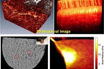
Non-invasive in vivo tissue imaging with functional optical coherence tomography (OCT)

Four 3D structual images of tissue of tissue imaging with functional optical coherence tomography

At a glance

Completed
Award date
July 2017 - November 2018
Contract amount
£49,947
Contractor(s)

R

  • Refinement

Overview

Tissue assessment is important in a variety of different applications, including clinical diagnosis and treatment, pharmaceutical and chemical development, and tissue engineering. The most basic and important parameter assessed is tissue structural information which indicates change on a cellular level. However, structural information alone is not enough to fully understand the tissue physiology.

Researchers from the University of Dundee have developed functional optical coherence tomography (OCT) to enable high quality, ultrafast, qualitative and quantitative characterisation of both tissue structure and function in vivo. The technique has the potential to both reduce and refine animal use, as long-term serial tissue monitoring in small animals is possible, and more invasive tissue assessment is avoided.  There is also the possibility to use OCT on biomimetic tissue models, enabling more information to be generated in these models without destroying the tissue, potentially opening up new opportunities for these non-animal models to replace some in vivo work.

They were seeking partners to apply this technique to their research, particularly in the areas of tissue engineering, diabetes or wound healing. Through CRACK IT Solutions, Professor Huang and her team are now working with Thea Pharmaceuticals and Keele University to explore the potential use of OCT in monitoring corneal wound healing.  Multiple animal models for corneal wound healing are currently used including rodent, rabbit and dog models. These can be extremely painful due to the high density of the nerves in the eye and can result in infections. Thea Pharmaceuticals have recently developed a new drug to promote corneal ulcer healing. OCT will be used to monitor the effect of this drug on tissue repair and regeneration in a human corneal tissue model, providing an excellent example of replacing the use of animal models for assessing drug effects on human tissue analogues.

Full details about this CRACK IT Solution and the output of the funded project can be found on the CRACK IT website.

Publications